Skip main navigation

Military Health System

Clear Your Browser Cache

This website has recently undergone changes. Users finding unexpected concerns may care to clear their browser's cache to ensure a seamless experience.

Images in Health Surveillance: Skin Rashes in Children Due to Infectious Causes

Image of Images in Health Surveillance: Skin Rashes in Children Due to Infectious Causes. Images in Health Surveillance: Skin Rashes in Children Due to Infectious Causes

Background

In this issue of the MSMR, an overview of the incidence of scarlet fever in Military Health System beneficiaries under 17 years of age is presented.1 The following provides a brief comparison of the characteristics of scarlet fever to other erythematous rashes associated with infectious diseases.

Scarlet fever

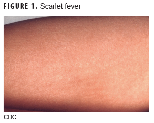
Scarlet fever (Figure 1) is caused by group A beta-hemolytic streptococcus bacteria.2 The incubation period is generally 2–5 days with prodromal symptoms of fever, sore throat, abdominal pain, and vomiting for 12–48 hours. The rash typically starts on the face or neck and rapidly spreads to the whole body, including the hands and feet, and is characterized as red, maculopapular, rough lesions commonly referred to as a sandpaper rash. Areas of skin folding—such as the groin, armpits, elbows, and knees—will typically develop a darker redness than other areas with the rash. The duration of the rash is variable from a few days to about 1 week and may be followed by desquamation or peeling of the skin for 1–3 weeks. Associated clinical findings include tonsillitis with cervical lymphadenopathy and a strawberry tongue.2,3

Measles

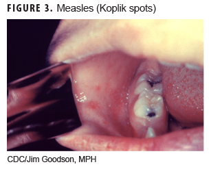
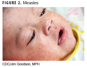
The rubeola virus is the etiologic agent for this infection (Figure 2). After an incubation period of 8–12 days, prodromal symptoms of fever, cough, coryza, and conjunctivitis begin.4 The rash appears 3–4 days after prodromal symptoms and begins around the ears and hairline on the face and spreads downward, covering the face, trunk, and arms by the second day. Initially the rash is red and maculopapular and becomes confluent by day 3. The rash typically lasts about 5 days and then fades in the same sequence as it appeared. Desquamation or peeling of the skin can follow the rash but does not occur on the palms or soles. The rash is not pruritic. Associated clinical findings include prodromal signs and Koplik spots (Figure 3) in the oral mucosa (white pinpoint-sized lesions with a reddened base).2,4,5

Varicella (chickenpox)

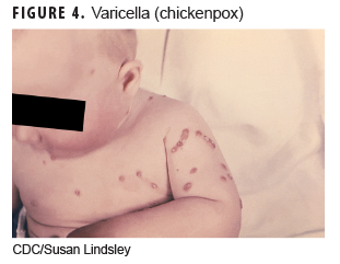
This disease (Figure 4) is caused by the initial infection with varicella-zoster virus. The incubation period is 14–16 days with a prodromal period of 0–2 days including fever, headache, malaise, abdominal pain, and decreased appetite. The rash may start on the chest, back, and face and then spreads over the whole body and is characterized by progression from vesicles in a teardrop shape that then crust and scab over. Patients typically have different stages of the rash on the body when examined. Usually within 24–48 hours, the vesicles progress to the crusting stage. All lesions progress to crusting by 5–10 days. The rash is very itchy. Associated clinical findings include high fever and lymphadenopathy.

Rubella (German measles)

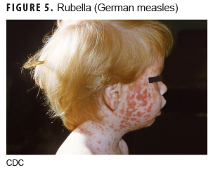
Rubella (Figure 5) is caused by the rubella virus and has an incubation period of 16–18 days with a prodromal period of 1–5 days before rash development, which consists of low-grade fever (less than 101°F), headache, conjunctivitis, malaise, lymphadenopathy, cough, and rhinorrhea.6 The rash typically starts on the face and spreads to the extremities over the next 48 hours and appears as small, fine, maculopapular, pink lesions that tend not to coalesce as the measles rash does. Associated clinical findings include distinctive lymphadenopathy including posterior cervical, suboccipital, and posterior auricular nodes.2,5

Erythema infectiosum

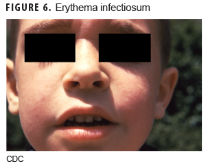
This illness (Figure 6) is caused by human parvovirus B19. The incubation period is 1–2 weeks, and a prodromal period lasts 2–5 days before the rash appears and consists of low-grade fever, coryza, headache, malaise, nausea, and diarrhea.7 The first stage of the rash usually begins on the cheeks as a solid bright red eruption with circumoral pallor, giving it a "slapped cheek" appearance. Over the next 1–4 days, the second stage of the rash develops, which is characterized by a maculopapular rash spreading to the trunk and extremities. If central clearing of the rash occurs, it will have a lacelike, reticular pattern. The rash is pruritic and typically fades over 1–3 weeks. Associated clinical conditions include arthropathy; transient aplastic crisis; chronic red cell aplasia; hydrops fetalis; and papular, pruritic eruptions on the hands and feet ("gloves and socks" syndrome).2,5

Roseola (exanthema subitum)

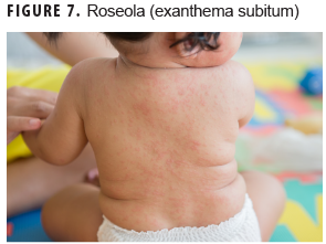
Human herpesvirus 6 (HHV-6) is the most common cause of this illness (Figure 7), but other viral causes include HHV-7, enteroviruses, adenoviruses, and parainfluenza type 1. The incubation period is 5–15 days, and a prodromal period consists of high fevers (104–105°F) for 3–4 days.8 Febrile convulsions may occur in young children. The rash appears as the fever resolves and begins on the chest and abdomen and spreads to the face and extremities and appears as small, separate, rose-pink, blanching, macular or maculopapular lesions. The rash typically resolves after 1–2 days without desquamation. The rash is not itchy. In addition to high fever, occipital adenopathy is a clinical finding along with the rash.2,5

Author affiliations: Department of Preventive Medicine and Biostatistics, Uniformed Services University of the Health Sciences, Bethesda, MD (Maj Sayers); Defense Health Agency, Armed Forces Health Surveillance Branch (Dr. Clark).

Disclaimer: The contents described in this publication are those of the authors and do not necessarily reflect official policy or position of Uniformed Services University of the Health Sciences, the Department of Defense, or Departments of the Army, Navy, or Air Force.

References

  1. Sayers DR, Bova ML, Clark LC. Brief report: Diagnoses of scarlet fever in Military Health System (MHS) beneficiaries under 17 years of age across the MHS and in England, 2013–2018. MSMR. 2020;27(2):26–27.
  2. Allmon A, Deane K, Martin KL. Common skin rashes in children. Am Fam Physician. 2015;92(3):211–216.
  3. Basetti S, Hodgson J, Rawson TM, Majeed A. Scarlet fever: a guide for general practitioners. London J Prim Care (Abingdon). 2017;9(5):77–79.
  4. Committee on Infectious Diseases, American Academy of Pediatrics. Measles. In: Red Book: 2018–2021 Report of the Committee on Infectious Diseases. 31st ed. Itasca, IL: American Academy of Pediatrics; 2018:537–550.
  5. Garcia JJG. Differential diagnosis of viral exanthemas. Open Vaccine J. 2010;3:65–68.
  6. Committee on Infectious Diseases, American Academy of Pediatrics. Rubella. In: Red Book: 2018–2021 Report of the Committee on Infectious Diseases. 31st ed. Itasca, IL: American Academy of Pediatrics; 2018;869–883.
  7. Committee on Infectious Diseases, American Academy of Pediatrics. Parvovirus B19 (Erythema Infectiosum, Fifth Disease). In: Red Book: 2018 Report of the Committee on Infectious Diseases. 31st ed. Itasca, IL: American Academy of Pediatrics; 2018:602–606.
  8. Committee on Infectious Diseases, American Academy of Pediatrics. Human herpesvirus 6 (including roseola) and 7. In: Red Book: 2018–2021 Report of the Committee on Infectious Diseases. 31st ed. Itasca, IL: American Academy of Pediatrics; 2018:454–457.
Scarlet fever

MeaslesMeasles (Koplik spots)Varicella (chickenpox)Rubella (German measles)Erythema infectiosumRoseola (exanthema subitum)

You also may be interested in...

Article
Apr 1, 2019

Modeling Lyme Disease Host Animal Habitat Suitability, West Point, New York

A deer basks in the morning sun at Joint Base San Antonio-Fort Sam Houston, Texas.  (Photo Courtesy: U.S. Air Force)

As the most frequently reported vector-borne disease among active component U.S. service members, with an incidence rate of 16 cases per 100,000 person-years in 2011, Lyme disease poses both a challenge to health care providers in the Military Health System and a threat to military readiness. Spread through the bite of an infected blacklegged tick, ...

Article
Mar 1, 2019

Sexually Transmitted Infections, Active Component, U.S. Armed Forces, 2010–2018

Anopheles merus

This report summarizes incidence rates of the 5 most common sexually transmitted infections (STIs) among active component service members of the U.S. Armed Forces during 2010–2018. Infections with chlamydia were the most common, followed in decreasing order of frequency by infections with genital human papillomavirus (HPV), gonorrhea, genital herpes ...

Article
Mar 1, 2019

Vasectomy and Vasectomy Reversals, Active Component, U.S. Armed Forces, 2000–2017

Sperm is the male reproductive cell  Photo: iStock

During 2000–2017, a total of 170,878 active component service members underwent a first-occurring vasectomy, for a crude overall incidence rate of 8.6 cases per 1,000 person-years (p-yrs). Among the men who underwent incident vasectomy, 2.2% had another vasectomy performed during the surveillance period. Compared to their respective counterparts, the ...

Article
Mar 1, 2019

Brief Report: Male Infertility, Active Component, U.S. Armed Forces, 2013–2017

Sperm is the male reproductive cell  Photo: iStock

Infertility, defined as the inability to achieve a successful pregnancy after 1 year or more of unprotected sexual intercourse or therapeutic donor insemination, affects approximately 15% of all couples. Male infertility is diagnosed when, after testing both partners, reproductive problems have been found in the male. A male factor contributes in part ...

Article
Mar 1, 2019

Testosterone Replacement Therapy Use Among Active Component Service Men, 2017

Image of Marines carrying a wooden log for physical fitness. Click to open a larger version of the image.

This analysis summarizes the prevalence of testosterone replacement therapy (TRT) during 2017 among active component service men by demographic and military characteristics. This analysis also determines the percentage of those receiving TRT in 2017 who had an indication for receiving TRT using the 2018 American Urological Association (AUA) clinical ...

Article
Feb 1, 2019

Update: Malaria, U.S. Armed Forces, 2018

Anopheles merus

Malaria infection remains an important health threat to U.S. service mem­bers who are located in endemic areas because of long-term duty assign­ments, participation in shorter-term contingency operations, or personal travel. In 2018, a total of 58 service members were diagnosed with or reported to have malaria. This represents a 65.7% increase from ...

Article
Feb 1, 2019

Update: Incidence of Glaucoma Diagnoses, Active Component, U.S. Armed Forces, 2013–2017

Glaucoma

Glaucoma is an eye disease that involves progressive optic nerve damage and vision loss, leading to blindness if undetected or untreated. This report describes an analysis using the Defense Medical Surveillance System to identify all active component service members with an incident diagnosis of glaucoma during the period between 2013 and 2017. The ...

Article
Feb 1, 2019

Outbreak of Acute Respiratory Illness Associated with Adenovirus Type 4 at the U.S. Naval Academy, 2016

Malaria case definition

Human adenoviruses (HAdVs) are known to cause respiratory illness outbreaks at basic military training (BMT) sites. HAdV type-4 and -7 vaccines are routinely administered at enlisted BMT sites, but not at military academies. During Aug.–Sept. 2016, U.S. Naval Academy clinical staff noted an increase in students presenting with acute respiratory ...

Article
Feb 1, 2019

Re-evaluation of the MSMR Case Definition for Incident Cases of Malaria

Anopheles merus

The MSMR has been publishing the results of surveillance studies of malaria since 1995. The standard MSMR case definition uses Medical Event Reports and records of hospitalizations in counting cases of malaria. This report summarizes the performance of the standard MSMR case definition in estimating incident cases of malaria from 2015 through 2017. ...

Article
Dec 1, 2018

Thyroid Disorders, Active Component, U.S. Armed Forces, 2008–2017

A U.S. naval officer listens through his stethoscope to hear his patient’s lungs at Camp Schwab in Okinawa, Japan in 2018. (Photo courtesy of U.S. Marine Corps) photo by Lance Cpl. Cameron Parks)

This analysis describes the incidence and prevalence of five thyroid disorders (goiter, thyrotoxicosis, primary/not otherwise specified [NOS] hypothyroidism, thyroiditis, and other disorders of the thyroid) among active component service members between 2008 and 2017. During the 10-year surveillance period, the most common incident thyroid disorder ...

Article
Dec 1, 2018

Incidence and Prevalence of the Metabolic Syndrome Using ICD-9 and ICD-10 Diagnostic Codes, Active Component, U.S. Armed Forces, 2002–2017

This report uses ICD-9 and ICD-10 codes (277.7 and E88.81, respectively) for the metabolic syndrome (MetS) to summarize trends in the incidence and prevalence of this condition among active component members of the U.S. Armed Forces between 2002 and 2017. During this period, the crude overall incidence rate of MetS was 7.5 cases per 100,000 person ...

Article
Dec 1, 2018

Adrenal Gland Disorders, Active Component, U.S. Armed Forces, 2002–2017

During 2002–2017, the most common incident adrenal gland disorder among male and female service members was adrenal insufficiency and the least common was adrenomedullary hyperfunction. Adrenal insufficiency was diagnosed among 267 females (crude overall incidence rate: 8.2 cases per 100,000 person-years [p-yrs]) and 729 males (3.9 per 100,000 p-yrs). ...

Fact Sheet
Mar 30, 2017

Demographic and Military Traits of Service Members Diagnosed as Traumatic Brain Injury Cases

.PDF | 283.00 KB

This fact sheet provides details on the demographic and military traits of service members diagnosed as traumatic brain injury (TBI) cases during a 16-year surveillance period from 2001 through 2016, a total of 276,858 active component service members received first-time diagnoses of TBI - a structural alteration of the brain or physiological ...

Fact Sheet
Mar 30, 2017

Rhabdomyolysis by Location, Active Component, U.S. Armed Forces, 2012-2016 Fact Sheet

.PDF | 313.80 KB

This fact sheet provides details on Rhabdomyolysis by location for active component, U.S. Armed Forces during a five-year surveillance period from 2012 through 2016. The medical treatment facilities at nine installations diagnosed at least 50 cases each and, together approximately half (49.9%) of all diagnosed cases.

Fact Sheet
Mar 30, 2017

Heat Illnesses by Location, Active Component, U.S. Armed Forces, 2012-2016 Fact Sheet

.PDF | 267.04 KB

This fact sheet provides details on heat illnesses by location during a five-year surveillance period from 2012 through 2016. 11,967 heat-related illnesses were diagnosed at more than 250 military installations and geographic locations worldwide. Three Army Installations accounted for close to one-third of all heat illnesses during the period.

Skip subpage navigation
Refine your search
Last Updated: July 11, 2023
Follow us on Instagram Follow us on LinkedIn Follow us on Facebook Follow us on X Follow us on YouTube Sign up on GovDelivery